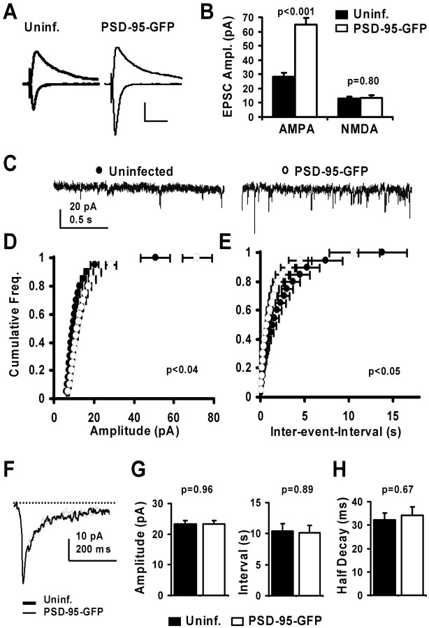
Figure 1.
Expression of PSD-95–GFP specifically potentiates AMPA-mediated EPSCs. A, Synaptic currents recorded simultaneously from nearby pyramidal neurons held at –60 and +40 mV, one uninfected and one expressing PSD-95–GFP. B, The AMPA component of EPSCs was significantly increased from 28.5 ± 2.5 to 64.8 ± 4.9 pA (control and infected neurons, respectively; n = 51), where as the late NMDA component was not altered (13.1±1.3 and 13.5±1.8 pA, control and infected neurons, respectively; n = 33). Calibration: 20 pA, 50 msec. C, Miniature EPSCs recorded at –60 mV from a pair of CA1 neurons. D, E, Cumulative histograms of amplitude and interevent interval for AMPA minis (n = 6 cells/group). The mini amplitude increases, whereas the interevent-interval decreases significantly (t test for each binned data point). F, Overlaid average miniature NMDA currents recorded from a pair of CA1 neurons recorded at –60 mV in 0 mm Mg 2+ and 5 μm CNQX. G, Plots of amplitude (23.3 ± 1.1 and 23.4 ± 1.0 pA, control and infected, respectively) and interevent interval (10.4 ± 1.2 and 10.1 ± 1.2 sec, control and infected, respectively) for average NMDA minis indicate no significant change (n = 10 cells/group; t test). H, Time to half decay of average NMDA minis (32.3 ± 2.9 and 34.3 ± 3.6 msec, control and PSD-95–GFP-expressing neurons, respectively) also shows no difference (n = 10 cells/group; t test).

