
Figure 2.
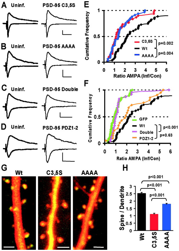
Membrane localization and PDZ interactions are necessary and sufficient for potentiation of AMPA currents. A–D, Synaptic currents measured in paired recordings from nearby control and infected neurons expressing mutant forms of PSD-95–GFP at –60 and +40 mV. Calibration: 20 pA, 50 msec. E, F, Cumulative distributions of AMPA-EPSC ratios (AMPAInf/AMPAUninf) in paired recordings are used to compare AMPA current potentiation by different mutants (p values from KS tests). E, The small potentiating effect of PSD-95-C3, 5S (n = 30) and PSD-95-AAAA (n = 33) was highly significantly different from wt PSD-95 (p = 0.002 and p = 0.004, respectively). F, Like GFP (n = 47), PSD-95-Double (n = 24) did not potentiate AMPA currents and was significantly different from wt PSD-95 (p < 0.001), whereas PSD-95–PDZ1–2 (n = 21) potentiated AMPA–EPSCs similar to wt-PSD-95 (p = 0.63). G, Subcellular distribution of mutant forms of PSD-95 compared with wt PSD-95–GFP. Dual-wavelength two-photon images of CA1 pyramidal neurons in slice cultures (9 d in vitro) coexpressing DsR-T1 and GFP-tagged PSD-95 constructs for ∼40–48 hr. Scale bars, 2μm. H, Spine/dendrite ratio shows that wt-PSD-95 is strongly accumulated in spines (ratio, 2.51 ± 0.07; n = 312 spines), C3, 5S is passively distributed (ratio, 1.11 ± 0.06; n = 285 spines), and the AAAA mutant is accumulated in spines (ratio, 1.78 ± 0.06; n = 323 spines), but less than wt (from three neurons each; KS tests).