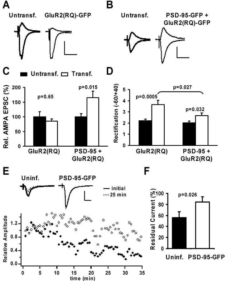
Figure 4.
PSD-95 does not affect the constitutive pathway of AMPAR delivery. A, B, AMPA-EPSCs recorded simultaneously from nearby CA1 neurons at –60 and +40 mV in 100μm APV. Calibration: 30 pA, 40 msec. A, Expression of GluR2(R607Q)–GFP did not alter AMPA-EPSC amplitude, but rectification was increased. B, PSD-95–GFP was coexpressed with GluR2(RQ)–GFP. AMPA-EPSC amplitude was strongly increased, but rectification was only slightly increased. C, Relative AMPA-EPSCs were not significantly different in control versus GluR2(RQ)-transfected neurons (100 ± 17.2 and 84.9 ± 7.6%; n = 15), but significantly increased in control versus neurons coexpressing PSD-95–GFP and GluR2(RQ)–GFP (100±11.2 vs 164.8± 22.3%; n = 17). D, Rectification was strongly increased in control versus GluR2(RQ)–GFP-transfected neurons (2.2 ± 0.2, n = 16 vs 3.7 ± 0.4, n = 13; t test) and slightly increased in control versus PSD-95–GFP and GluR2(RQ)–GFP-expressing neurons (2.0 ± 0.2, n = 16 vs 2.7 ± 0.2, n = 16; t test). Importantly, rectification in neurons coexpressing PSD-95 and GluR2(RQ) was significantly smaller than in neurons expressing GluR2(RQ) alone (t test). E, Changes in synaptic AMPA currents recorded during infusion of peptide pep2m (2 mm) into two nearby neurons. EPSC amplitudes decreased in the uninfected neuron, but there was little change in a neuron expressing PSD-95–GFP. Calibration: 40 pA, 20 msec. F, The residual AMPA-EPSC 25 min after infusion of pep2m is significantly smaller in control than infected neurons (55.8 ± 10.7 vs 84 ± 9.5% of initial value; n = 11; t test).

