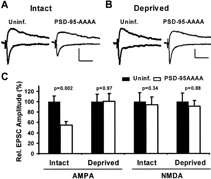
Figure 8.
Expression of dominant negative PSD-95 blocks experience-driven AMPAR delivery to synapses in barrel cortex. A, B, EPSCs evoked in layer IV and recorded simultaneously from nearby layer II/III pyramidal neurons held at –60 and +40 mV. Calibration: 10 pA, 40 msec. A, In intact whisker animals, the AMPAR- but not the NMDAR-mediated EPSC was depressed in a neuron expressing PDS-95AAAA–GFP. B, In deprived animals, there was no change in AMPA or NMDA currents in the control versus the infected neuron. C, Summary of relative changes in AMPAR- and NMDAR-mediated EPSCs. The AMPA component was significantly depressed by PSD-95AAAA when whiskers were intact (100 ± 11.1 and 54.5 ± 7.0%, control and infected; n = 18), but no change was seen during contralateral deprivation (100 ± 14.3 and 100.3 ± 15.2%, control and infected; n = 20). In all cases, NMDA currents were not significantly changed (Intact, 100 ± 16.7 and 93.5 ± 15.1%, control and infected, n = 14; Deprived, 100 ± 16.4 and 90.9 ± 11.4%, control and infected, n = 16).

