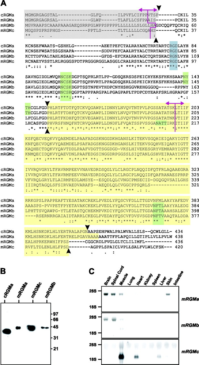
Figure 1.

Characterization of the murine RGM protein family. A, Protein sequence alignment of cRGMa, mRGMa, mRGMb, and mRGMc. Asterisks indicate identical amino acids; pink lines, intron–exon junctions; the gray box, predicted signal peptides; the blue box, potential integrin binding sites (RGD); green boxes, predicted N-glycosylation sites; the yellow box, mature C-terminal RGM fragments after full proteolytic cleavage and C-terminal GPI anchor addition (proteolytic cleavage sites indicated by arrowheads). B, Western blot analysis of supernatant collected from COS-7 cells transfected with C-terminally truncated histidine–Myc-labeled cRGMa, mRGMa, mRGMc, and mRGMb detected with an anti-Myc antibody. Molecular weight standards in kilodaltons are indicated on the right. C, Northern blot analysis on total RNA from a variety of P3 mouse tissues as indicated, using mRGMa, mRGMb, and mRGMc as probes.
