
Figure 8.
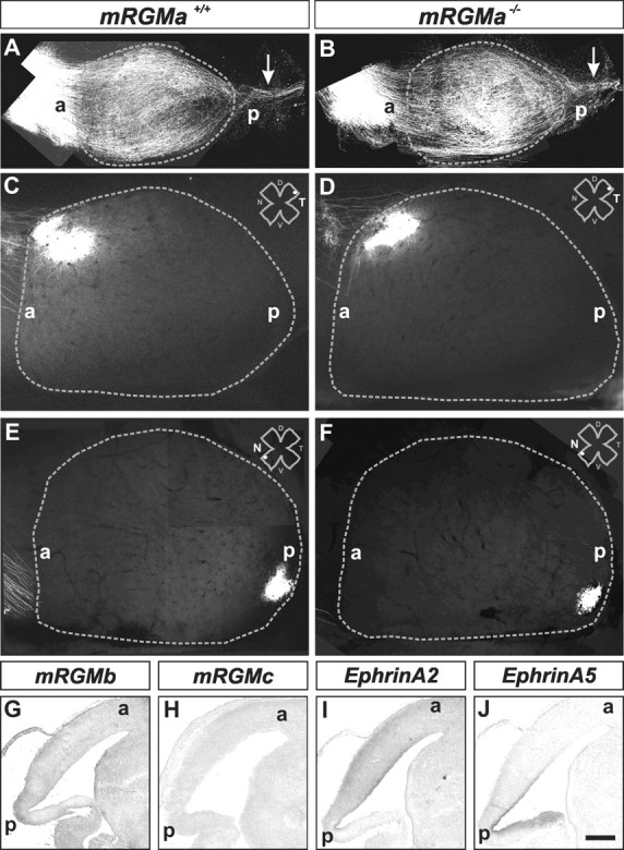
Lack of retinocollicular projection phenotype in mRGMa mutant mice. A, B, Dorsal views of superior and inferior colliculi from mRGMa+/+ (A) and mRGMa–/– (B) mice after anterograde Dil labeling from the retina at P0 to label many RGC axons. The gray dashed line outlines the superior colliculus; arrows point to RGC axons projecting to the inferior colliculus. C–F, Anterograde Dil labeling from the temporal (C, D) or nasal (E, F) retina to the superior colliculus (outlined by gray dashed lines) after focal Dil injection into the retina of mRGMa+/+ (C, E) and mRGMa–/– (D, F) mice at P10 (injection point in the retina indicated by the white dot the top right corner: T, Temporal; N, nasal; D, dorsal; V, ventral). G–J, Expression of mRGMb (G), mRGMc (H), mEphrinA2(I), and mEphrinA5 (J) in superior colliculi of E16.5 sagittal brain sections from mRGMa–/– mice detected by in situ hybridization. a, Anterior; p, posterior. Scale bar: (in J) A, B, 0.5 mm; C–F, 0.33 mm; G–J, 0.3 mm.