
Figure 1.
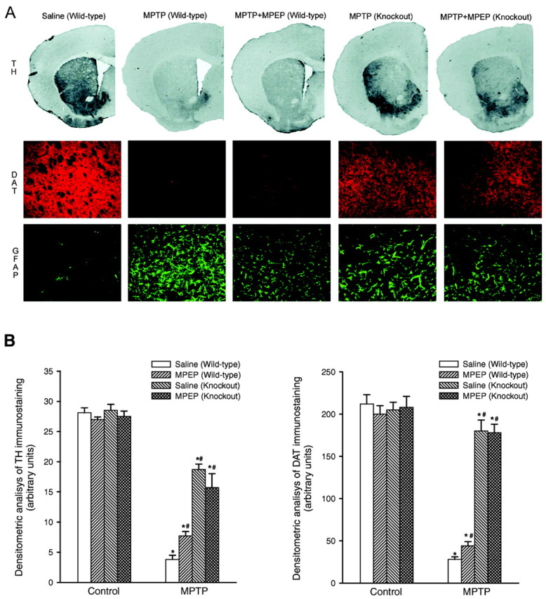
A, Immunohistochemical analysis of TH, DAT, and GFAP in the corpus striatum of wild-type and mGlu5 knock-out mice injected with saline or MPTP (20 mg/kg, i.p., injected 4 times with a 2 hr interval) alone or combined with MPEP (5 mg/kg, i.p., injected 4 times, 30 min before each saline or MPTP injection). Scale bar, 50 μm. B, Densitometric analysis of striatal TH and DAT immunostaining was performed on comparable sections from five to six mice. *p < 0.05 (one-way ANOVA plus Fisher's PLSD) versus the respective control mice treated with saline. #p < 0.05 (one-way ANOVA plus Fisher's PLSD) versus wild-type mice treated with MPTP and saline. Note that the lateral ventricle is not visible in the two representative sections of mGlu5 knock-out mice. This particular feature was observed in most of the sections from knock-out mice and was apparently attributable to a hypertrophy of the choroid plexus.