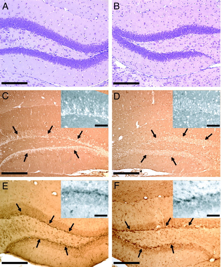
Figure 5.

Immunohistochemical analysis of the two most highly differentially expressed genes in the dentate gyrus, the cellular isoform of prion protein (PrPc) and the Sos2 protein. A, B, Nissl-stained sections of the dentate gyrus from an injured brain to illustrate cellular architecture, both contralateral and ipsilateral to the site of fluid percussion injury, respectively. C, A clear absence of PrPc-immunopositive cells is observed surrounding the subgranular layer of the dentate gyrus of the hippocampus contralateral to the site of injury, whereas such laminar organization (best illustrated by higher magnification insets) is not seen in the ipsilateral dentate gyrus, and many more cells immunolabel positively for PrPc (D). E, F, Sos2-immunopositive cells were found bilaterally throughout the subgranular layer of the dentate gyrus after TBI, although immunolabeling is consistently more intense in the subgranular layer ipsilateral to the site of injury (F). Scale bars: larger images, 200 μm; inset photomicrographs, 50 μm.
