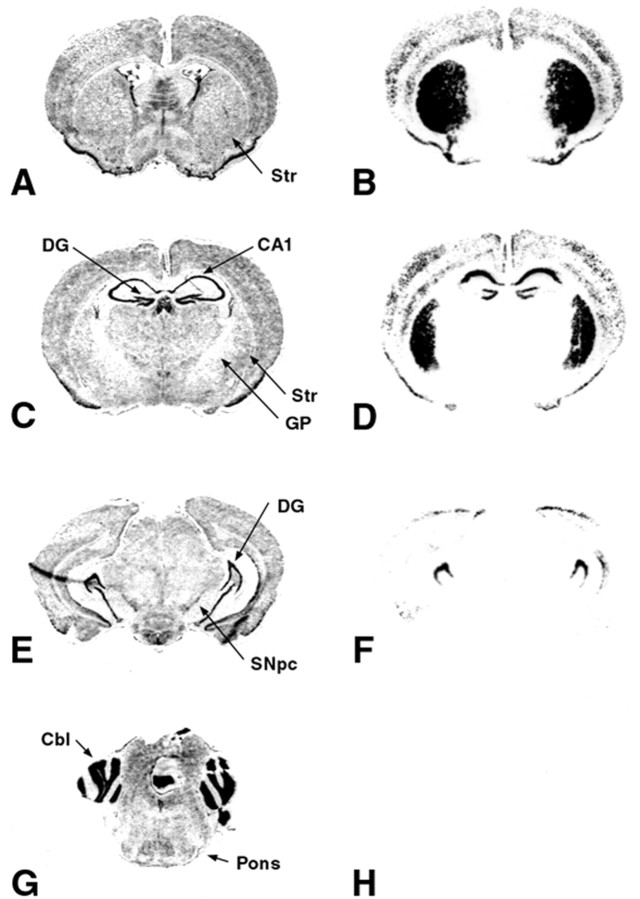
Figure 3.
Regional patterns of LacZ expression in line 14 double transgenic mice. A, C, E, G, Nissl-stained coronal sections adjacent to the corresponding LacZ-stained sections shown in B, D, F, H. A, B, Correspond to anterior Paxinos and Franklin (2001) plane 4.06 (interaural); C, D, plane 2.46; E, F, plane 0.88; G, H, most caudal, plane –1.22. There is robust LacZ staining in striatum (Str), the CA1 region and the dentate gyrus (DG) of the hippocampus, and the cortex. There is no staining in the globus pallidus (GP), the SNpc, or the cerebellum (Cbl). Thus, LacZ expression is observed only in target regions of the mesencephalic dopaminergic projection.

