
Figure 3.
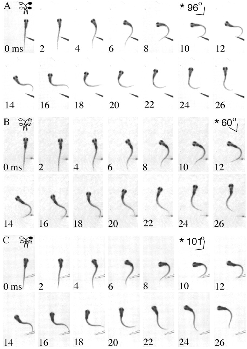
Time series illustrating postablation startles of larval zebrafish. The angle of the head in frames indicated by an asterisk shows the peak angle at the end of the initial lateral bend compared with the position of the body just before movement (0 msec). Fish are startled with a fine glass probe, and their behavior is recorded with high-speed video at 1000 Hz. A, A time series of images illustrating startle in a fish with both r2 and r4 M-cells. The fish turns ∼96° at 10 msec. The initial body bend is followed by continued swimming movements reflective of a typical larval zebrafish startle. B, Startle after both the r2 and r4 Mauthner cells are lesioned. The fish turns away from the tactile stimulus, forming a peak angle of 60° at 12 msec. The startle is followed by rhythmic swimming. C, The same pattern of startle behavior is seen in fish after the r4 Mauthner cell is ablated with a turn away from the stimulus. However, the angle at the end of the initial bend is higher than that seen with both cells lesioned, 101° at 10 msec. The startle is followed by rhythmic swimming.