
Figure 3.
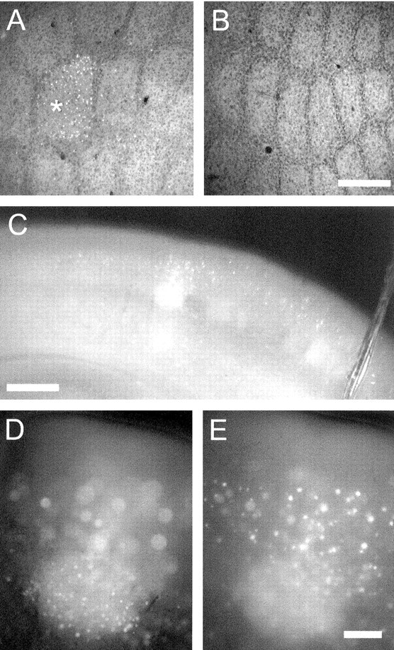
Sensory-induced expression of fosGFP in living brain tissue. All but a single large facial vibrissa, whisker D1, were removed by plucking from a fosGFP transgenic mouse (line 1-3, line 4-1, or line 5-1), aged ∼4 weeks. The animal was returned to its home cage for 24 hr before brain slices were prepared. A, Fixed and flattened section of cortex from the deprived hemisphere, showing fosGFP fluorescence in cells from layer IV of the spared whisker barrel, D1 (indicated by asterisk). B, Fixed and flattened cortex from the contralateral, unplucked hemisphere showing no fosGFP signal in the barrels. Scale bar, 100 μm. C, Low-magnification view of a coronal section of cortex, showing a single medial barrel that corresponds to the D1 whisker showing strong GFP fluorescence in living tissue. Scale bar, 200 μm. D, High-magnification view of C, with layer IV of the spared barrel in focus. Note the sharp edges delineated by fosGFP expression, corresponding to the margins of the spared barrel. E, High-magnification view of C, with layer II-III of the spared barrel in focus. Scale bar: B, C, 100 μm.