
Figure 7.
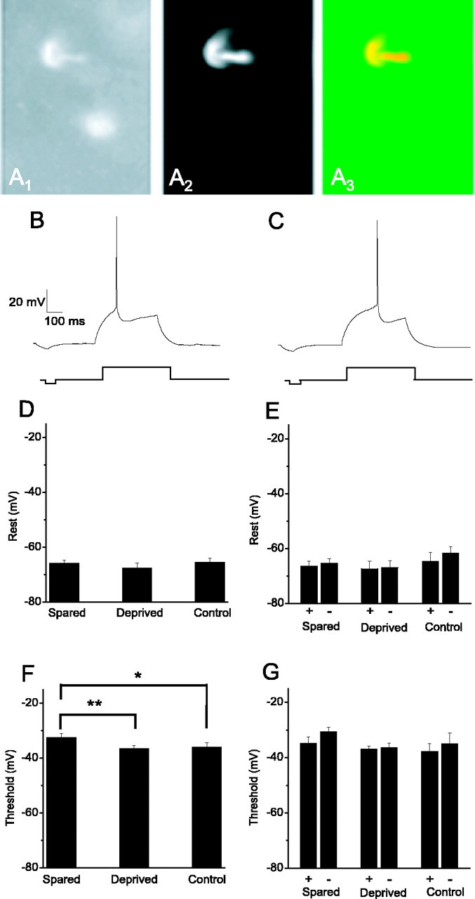
Threshold for action potential generation is raised in the spared barrel. A, Example of fosGFP+ cell from layer II-III of barrel cortex that was targeted for whole-cell voltage-clamp recording. A1, fosGFP+ nuclei. A2, Patch solution contained the red fluorescent dye Alexa-568 (10 μm) to fill the targeted cell during recording. A3, Merged picture of A and B shows that the Alexa-filled cell has a fosGFP+ nucleus. Scale bar, 30 μm. B, C, The minimal current to elicit an action potential in fosGFP+ (B) or fosGFP- (C) neurons under control conditions was determined, and the spike threshold was determined by averaging the inflection point from at least two rheobase traces. D, Resting potential from neurons in spared (n = 30), deprived (n = 22), or control (n = 28) tissue was unaltered. E, Data for Vrest of fosGFP+ and fosGFP- neurons for each experimental condition (spared, n = 13+, 17-; deprived, n = 8+, 13-; control, n = 8+, 9-). F, The average threshold for neurons in the spared barrel (n = 28) was altered compared with neurons from whisker-deprived barrels (**p < 0.05; n = 20) or control, unplucked animals (*p < 0.1; n = 15). G, Data for Vthreshold of fosGFP+ and fosGFP- neurons for each experimental condition (spared, n = 12+, 16-; deprived, n = 7+, 13-; control, n = 7+, 8-).