
Figure 1.
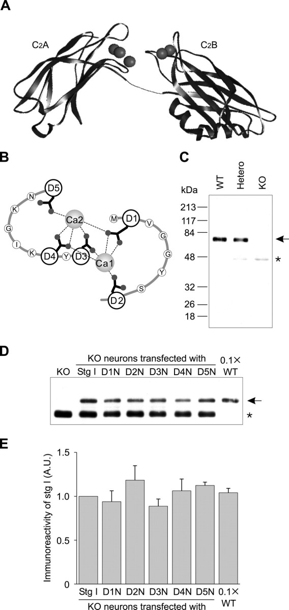
Expression of exogenous synaptotagmin I in the knock-out neurons. A, Vector NTI software (InfoMax, Frederick, MD) was used to generate structures of C2A (Shao et al., 1998) and C2B (Fernandez et al., 2001) domains in the presence of Ca2+ (spheres). The linker connecting the C2 domains was added manually. B, Ca2+-binding sites in the C2B domain of synaptotagmin I, modified from Fernandez et al. (2001). Two Ca2+ (Ca1 and Ca2) are coordinated by five Asp residues (D1 through D5) on two loops. C, Expression of synaptotagmin I in neurons from wild-type (WT), heterozygous (Hetero), or knock-out (KO) mice. Cultured hippocampal neurons (2 × 10³ cells) were subjected to immunoblotting. The migration position of synaptotagmin I is indicated by an arrow, and the truncated product weakly expressed in the synaptotagmin mutant is indicated by an asterisk. D, E, Expression of exogenous synaptotagmin I in the knock-out neurons. D, Knock-out neurons (2 × 10⁴ cells) either untransfected (KO) or transfected with either wild-type (Stg I) or mutated synaptotagmin I (D1N-D5N) were analyzed via immunoblotting. Wild-type neurons (WT; 2 × 10³ cells) were loaded as a control. E, Immunoreactivities were measured and normalized to wild-type synaptotagmin I expressed in the knock-out neurons. Data are the mean ± SEM from three experiments.