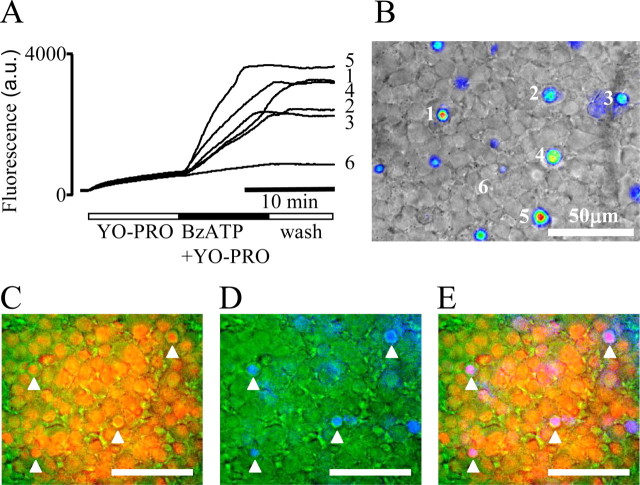
Figure 2.
BzATP-induced cell permeabilization in the rat inner retina. A shows the kinetics of YO-PRO-1 (1.5 μm) uptake in rat flat-mount retina exposed to prolonged stimulation with BzATP (10 min, 100 μm). B shows the pattern of YO-PRO-1 labeling in the GCL at the end of the BzATP stimulation. The marked regions correspond to the fluorescence traces shown in A. C, The cell nuclei were labeled with Hoechst 33342 dye (in red) before the experiment. D, YO-PRO-1 accumulation (in blue) was induced by prolonged stimulation with BzATP. E, Merged image of C and D; the white arrows point out the coincidence between YO-PRO-1 staining and labeled cell nuclei in the GCL. Scale bars, 50 μm. a.u., Arbitrary units.

