
Figure 4.
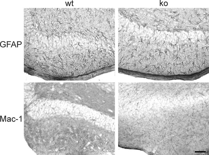
Astrocyte activation 8 d after entorhinal cortex lesion does not differ in wild-type and CXCR3 knock-out mice. Horizontal vibratome sections of hippocampus from wild-type mice (wt; left column) and CXCR3 knock-out mice (ko; right column) show GFAP-positive astrocytes (top panels) and Mac-1-positive microglia (bottom panels). No difference in the astrocytic activation could be observed between knock-out and wild-type animals. As described in Figure 2, microglial activation has already ceased at 8 days after lesion. Scale bar, 100 μm.