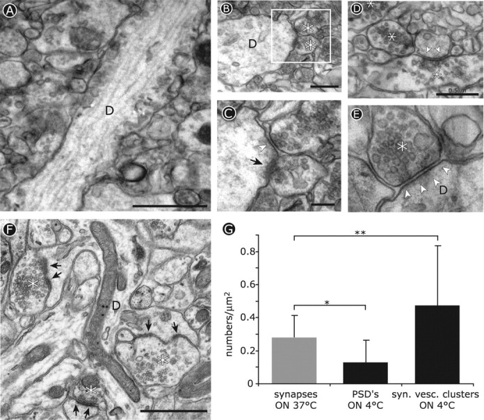
Figure 3.

Ultrastructural changes induced by cooled tissue slices to 4°C. A-E, Electron micrographs of dendrites in acutely cut brain slices from adult mice after incubation overnight at 4°C. The asterisks indicate presynaptic boutons, and arrows indicate the postsynaptic density. A, Longitudinal section of a dendrite (D) in area CA1 lacking dendritic spines on its surface. Scale bar, 1 μm. B, Synapses made directly onto the shafts of dendrites after cooling. Scale bar, 0.5 μm. C, Detail of the boxed area in B showing decreased staining of the postsynaptic density (arrowhead) at one synapse compared with its neighbor (arrow). Scale bar, 0.2 μm. D, Presynaptic specializations are maintained after cooling, whereas synaptic vesicle clusters are present at sites lacking a clear postsynaptic density (arrowheads). Scale bar, 0.5 μm. E, Synapses with marked presynaptic terminals and synaptic junctions often lack a clearly stained postsynaptic density after cooling. Scale bar, 0.2 μm. F, Ultrastructure of a control slice from the same tissue block maintained at 37°C overnight. The asterisks indicate presynaptic boutons, and the arrows indicate PSDs. Scale bar, 1 μm. G, Quantification of synaptic contact numbers in 37°C slices (gray bar) compared with indicators of synaptic structure (postsynaptic densities and synaptic vesicle clusters) in slices incubated at 4°C. *p < 0.025; **p < 0.05.
