
Figure 3.
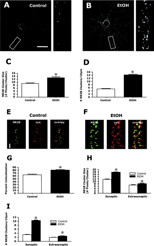
Chronic ethanol enhances the synaptic clustering of NR2B. A, B, Immunohistochemistry and confocal imaging of NR2B subunits revealed numerous small punctate clusters along dendrites similar to that observed with NR1. This clustering was greatly increased after exposure to ethanol (50 mm; 4 d) (compare A and B). Scale bar, 40 μm. C, D, Quantification of clustering indicated a significant increase in both cluster size (C) and density (D). The asterisk indicates a significant difference from control (p < 0.01, Student's t test; n = 90). E, G, Dual immunohistochemistry of NR2B (green) and synapsin (syn; red) revealed ∼65% colocalization of NR2B and synapsin clusters under control conditions (E, G). After ethanol exposure (50 mm; 4 d), this colocalization increased to ∼80% (F, G). The asterisk indicates significant difference from control (p < 0.01, Student's t test; n = 90). Scale bar, 5 μm. H, I, Analysis of clustering in the synaptic and extrasynaptic compartments revealed that ethanol treatment greatly increased the size (H) and density (I) of synaptic clusters, with only minor increases in extrasynaptic clusters. The asterisk indicates a significant difference from control (p < 0.01, Student's t test; n = 90).