
Figure 5.
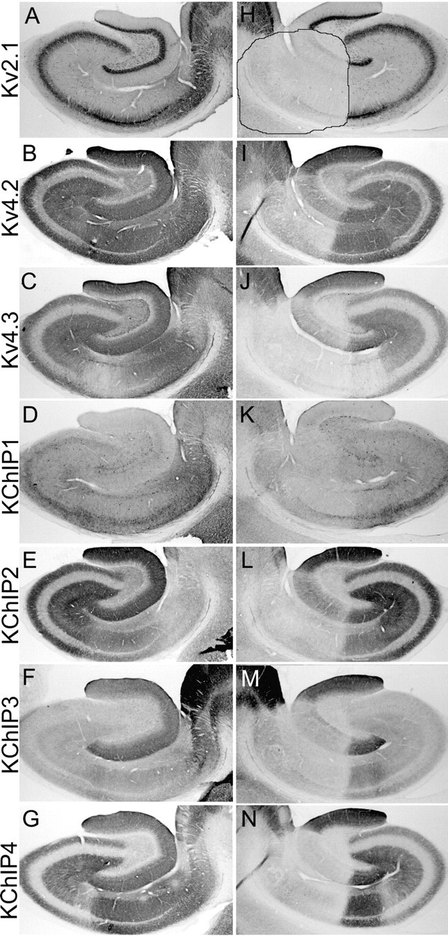
Effects of an ibotenic acid lesion on the distribution and density of Kv4 and KChIP immunoreactivity in the hippocampal formation. These photomicrographs show the pattern of immunoreactivity for the indicated subunits in the unoperated, control hemisphere (A-G) and operated hemisphere (H-N) of an animal that sustained a circumscribed unilateral ibotenic acid lesion. This lesion destroyed cells in the distal CA1 subfield, prosubiculum, and subiculum and also destroyed a central portion of the dentate gyrus. The entire CA3 and proximal CA1 subfield was spared by this lesion. The pattern of cell loss is clearly indicated by the loss of Kv2.1 immunoreactivity within the boundaries of the lesion (outlined in H). This lesion greatly reduced the density of Kv4.2 (compare B, I), Kv4.3 (compare C, J), and KChIP1-4 (compare D-G, K-N) immunoreactivity within the boundaries of the lesion, indicating that these subunits are localized to the cell bodies and/or dendrites of dentate granule cells and CA1 pyramidal cells or, in the case of Kv4.3 and KChIP1, to the cell bodies and dendrites of dentate and CA1 interneurons.