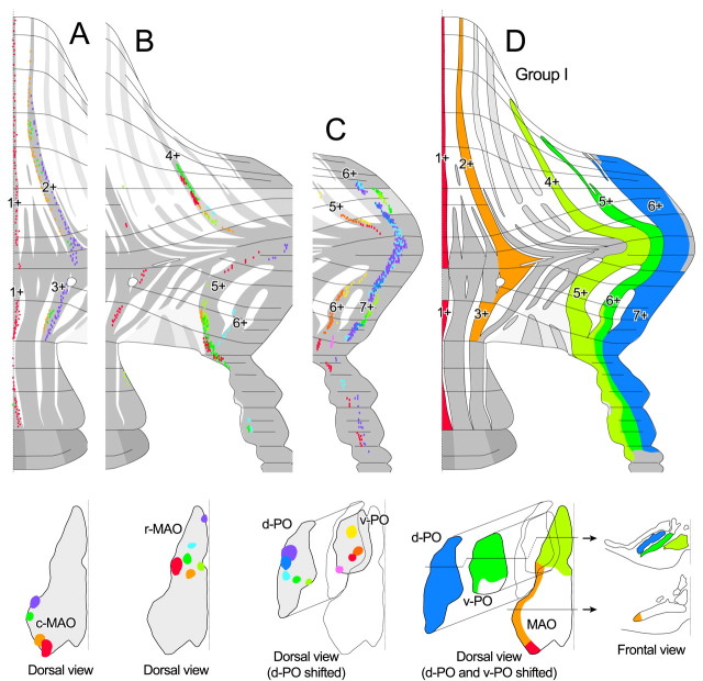
Figure 4.
Mapping of labeled climbing fibers and summarized topographic scheme of the olivocerebellar projection belonging to group I. This group consisted of projections from caudal subnucleus a and the intermediate and rostral subnucleus a of the caudal part of the MAO (c-MAO) to 1+//1+ and 2+//3+, respectively, from the rostral part of the MAO (r-MAO) to 4+//5+; from the ventral lamella of the PO (v-PO), except for the caudomedial part, to 5+//6+; and from the dorsal lamella of the PO (d-PO) to 6+//7+. A-C, Plots of labeled climbing fibers in 19 injections terminating in 1+//1+ and 2+//3+ (A), 4+//5+ (B), and 5+//6+ and 6+//7+ (C). Labeled climbing fibers in all of these experiments (A-C) were nearly aligned in a single narrow band in a pair of linked rostral and caudal aldolase C compartments, except for one case (B; red), in which some outside climbing fibers were seen. D, Putative olivocerebellar topography within group I.

