
Figure 2.
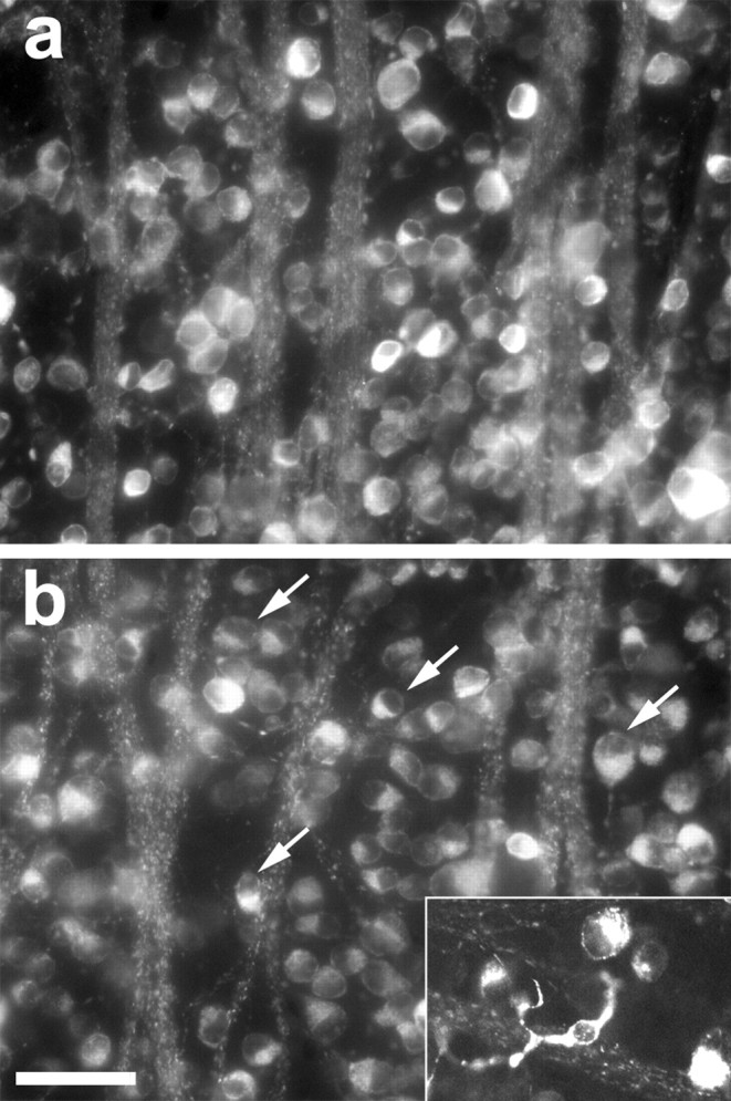
RGC survival after optic nerve injury. RGC morphology, survival, and phagocytosis were visualized in flat-mounted retinas dissected 4-7 d after axotomy; RGCs were retrogradely labeled with 4Di-10ASP 1 week before axotomy. RGCs appear normal 4 d after axotomy (a) but show eccentric nuclei after 6 d (b; arrows). Labeled microglia appear by day 7 (inset). Scale bar, 50 μm.