Figure 2.
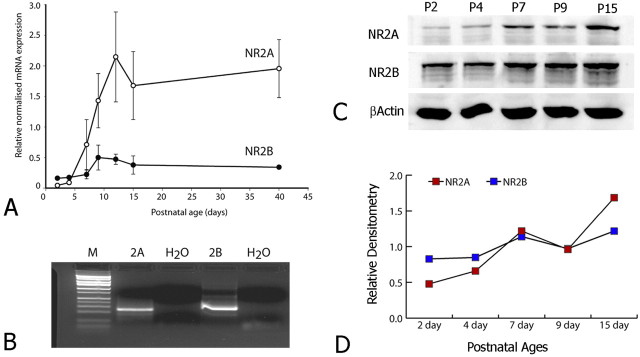
Assessment of NR2A and NR2B expression in somatosensory cortex by RT-PCR (A, B) and Western blot analysis (C, D). A, As judged by RT-PCR, the expression of both NR2A and NR2B mRNA is developmentally regulated. NR2A expression is extremely low at P2 but undergoes a rapid increase in expression from P4 to P12 and maintains this elevated expression through P40. In contrast, NR2B levels are greater than NR2A at P2 and undergo a slight transient increase from P7 to P15 but remain elevated through P40. B, RT-PCR amplifies a single band at the predicted size for both NR2A and NR2B. After real-time PCR analysis, samples were separated by gel electrophoresis and stained with ethidium bromide. H2O, Water only; M, DNA marker. C, NR2A and NR2B protein levels are also developmentally regulated. A Western blot analysis of NR2A, NR2B, and β-actin illustrates protein expression at various postnatal ages. The same amount of protein is loaded per lane. D, Densitometric measures of NR2A and NR2B levels obtained from Western blots are normalized to β-actin levels and expressed relative to levels of NR2B at P9. NR2A shows a dramatic increase in total cellular protein expression compared with NR2B, for which protein levels remain relatively constant throughout early postnatal development.

