Figure 3.

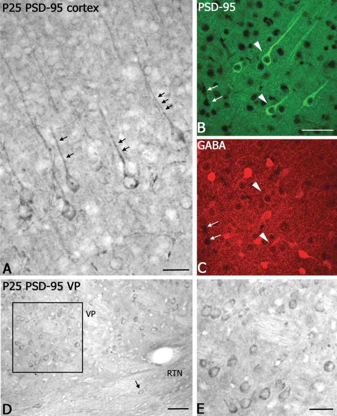
Immunocytochemical localization of PSD-95 in the cortex and thalamus. A, In somatosensory cortex, prominent PSD-95 immunoperoxidase-labeled pyramidal neurons are present in layer V. Note the strong immunolabeled apical dendrites (small black arrows). Scale bar, 50 μm. B, C, Laser confocal microscopic images of double immunofluorescent labeling for PSD-95 (B, green, fluorescein) and GABA (C, red, Cy5) show a lack of colocalization in the cortex (white arrowheads). As a point of reference, small white arrows indicate blood vessels in the section. Scale bar, 60 μm. D, Light micrograph showing PSD-95 immunoperoxidase labeled neurons in VP; a few labeled neurons are visible in RTN (small arrow). Scale bar, 100 μm. E, Higher-magnification image of PSD-95-immunolabeled neurons in VP nucleus (D, boxed region). Scale bar, 50 μm.
