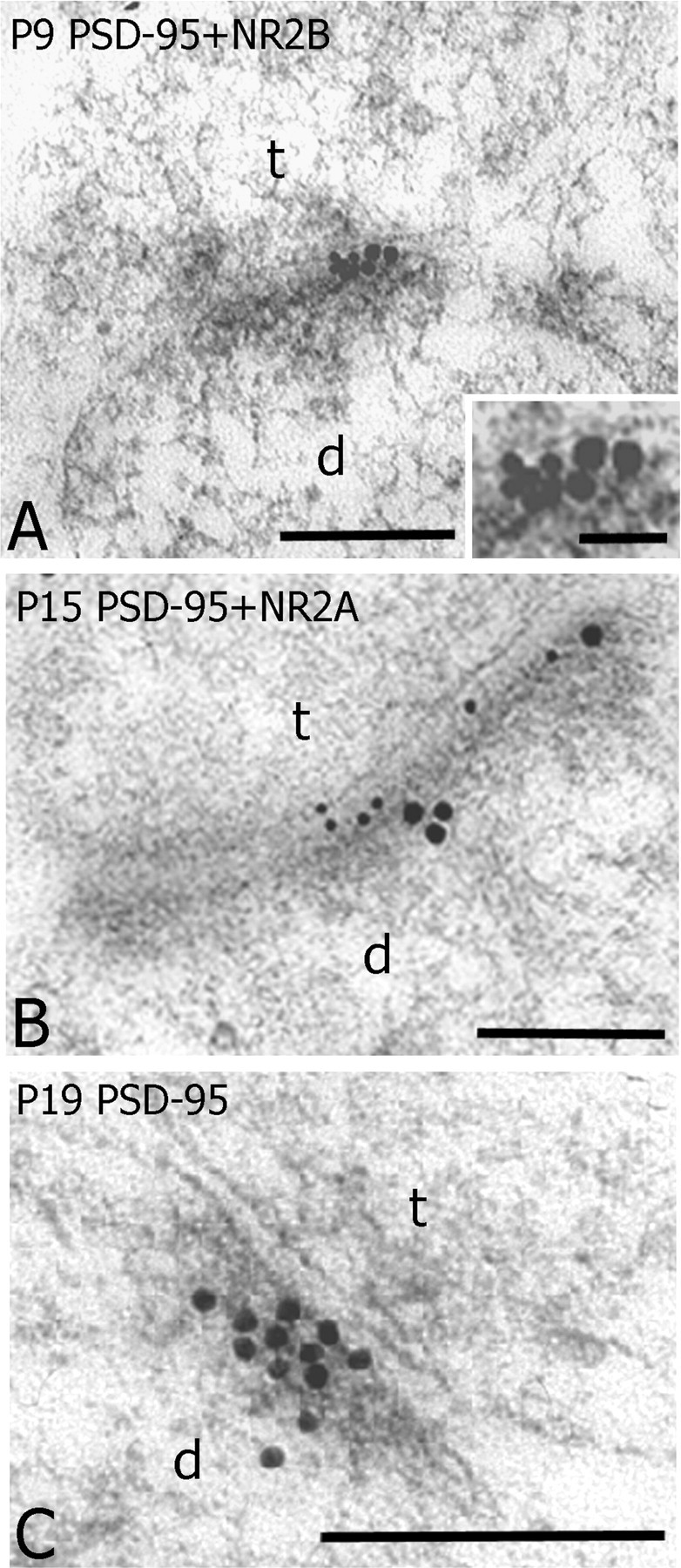
Figure 6.

Synaptic colocalization of PSD-95 and NR2 subunits at developing synapses in layer IV of somatosensory cortex. A, An electron micrograph showing PSD-95 (small particles, 10 nm) and NR2B (large particles, 15 nm) colocalized at the PSD of an asymmetrical synapse (t) contacting a dendritic profile (d) at P9. B, PSD-95 (10 nm particles) and NR2A (20 nm particles) also colocalized at a PSD profile of an asymmetrical synapse(t) contacting dendritic profile (d) at P15. C, A cluster of PSD-95 particles concentrated at the PSD profile of an asymmetrical synapse at P19. Scale bars: 0.2 μm; A, inset, 0.05 μm.
