Figure 7.
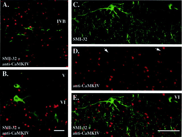
CaMKIV immunofluorescence signals do not colocalize with those of SMI-32, a maker of pyramidal neurons. A, B, Double immunofluorescence staining of SMI-32 (green) and a nonamplified CaMKIV signal (red) in layers IVB and VI of area V1 in a 5 d ME adult monkey. Scale bar, 50 μm. C, D, Immunofluorescence staining for SMI-32 (C, green), an amplified CaMKIV signal using the TSA method (D, red) in a second 5 d ME adult monkey. E, Digitally superimposed image of CaMKIV and SMI-32 staining showing a lack of spatial coincidence in the two signals. White arrows in D indicate the absence of CaMKIV immunostaining in SMI-32-positive neurons. Scale bar, 100 μm.

