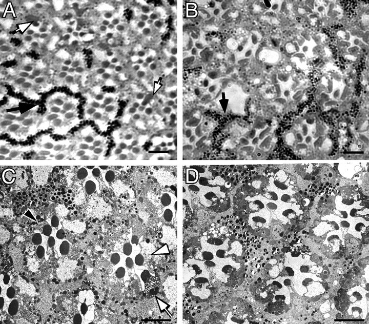
Figure 2.

The effect of ΔH99 [homozygous Df(H99) deletion] on wild-type and norpA photoreceptor degeneration. Light (A, B) and electron micrographs (C, D) of 3-d-old ΔH99 mosaic norpA+ and norpA mosaic ommatidia. The areas of the eye containing photoreceptors and surrounding pigment cells with pigment granules contain ΔH99+ cells, the area of the eye lacking pigment granules contain ΔH99 photoreceptor cells. A, norpA+ mosaics at 3 d. Ommatidia appear normal in both wild-type (pigmented) and ΔH99 (nonpigmented) regions of the mosaic norpA+ genotype. One region of pigment granules is identified by a black arrow. Several ΔH99 photoreceptors show increased cytoplasmic staining; two of these are identified by white arrows. B, norpA mosaics at 3 d. In the norpA mutant, a region of the retina with both ΔH99+ and ΔH99 photoreceptors show that all cells are indistinguishable from each other and are undergoing degeneration. One region of pigment granules is identified by a black arrow. The dispersion of the pigment granules (compare with A) is a result of the degeneration process. C, norpA+ mosaics at 3 d. The ΔH99+ cells, containing pigment granules (1 cell is marked by a black arrowhead) appear wild type, and the nonpigmented ΔH99 cells (1 cell is marked with a white arrowhead) also are wild type. One ΔH99 photoreceptor with cytoplasmic vacuoles (white arrow) identifies one of the abnormally stained cells noted in A. D, norpA mosaics at 3 d. The field contains an approximately equal number of norpA; ΔH99+ and norpA; ΔH99 photoreceptor cells, as assessed by the distribution of pigmented-nonpigmented pigment cells. Individual photoreceptors cannot be unambiguously assigned as H99+ or ΔH99 because both types of cells are undergoing degeneration. Scale bars, ∼5 μm.
