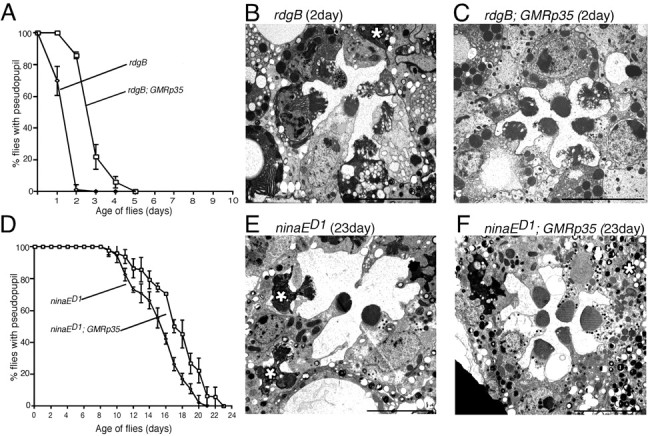
Figure 5.

The influence of P35 in the control of photoreceptor cell death in other retinal degeneration mutants. Flies were reared under constant light at 22°C. A, Retinal degeneration assessed by deep pseudopupil analysis in rdgB and rdgB; P35. B, C, Electron micrographs of 2-d-old rdgB (B) and rdgB; P35 (C) photoreceptors. The degenerated photoreceptor (marked by * in B) appears in the process of being phagocytosed. D, Retinal degeneration assessed by deep pseudopupil analysis in ninaED1 and ninaED1; P35. E, F, Electron micrographs of 23-d-old ninaED1 retina (E) and ninaED1; P35 (F). The degenerated photoreceptors (marked by * in E) are being phogocytosed. Scale bars, 5 μm.
