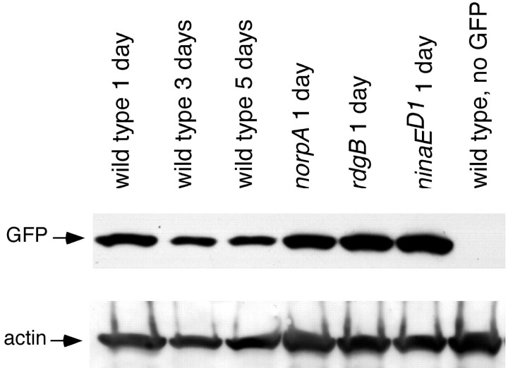
Figure 6.
Western blot analysis of GFP driven by the GMR promoter in adult and mutant photoreceptors. Flies were raised in 12 hr light/dark cycle at 22°C. Flies carrying both GMR-Gal4 and UAS-GFP were crossed into norpA, rdgB, and ninaED1/+ mutant backgrounds. The protein equivalent of one fly head is loaded into each lane for GFP detection (top row); the same blot was also probed for actin (bottom row) to examine loading differences. The GFP protein is detected in wild-type 1-, 3-, and 5-d-old heads (lanes 1-3). Similar levels of GFP protein are detected in 1-d-old norpA (lane 4), rdgB (lane 5), and ninaED1 (lane 6) heads.

