
Figure 1.
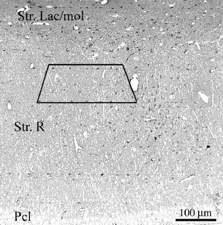
Light micrograph taken from a toluidine blue-stained, 2-μm-thick semithin section of the CA1 hippocampal subfield. The box labels the area from which the serial ultrathin sections were cut from each block. Str. Lac/mol, Stratum lacunosum moleculare; Str. R, stratum radiatum; Pcl, pyramidal cell layer.