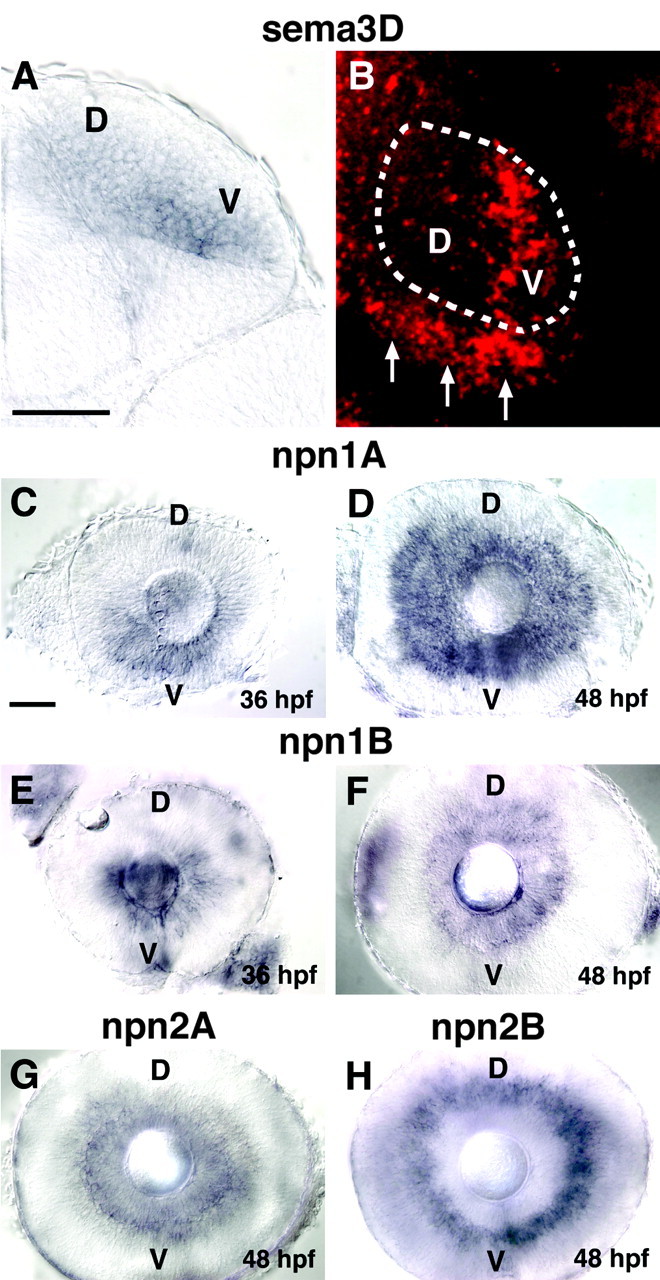
Figure 1.

Expression pattern of sema3D and neuropilins. A, Cross section through the midbrain (dorsal is up) of 48 hpf embryos showing sema3D in situ hybridization in the right tectum. sema3D mRNA is expressed more strongly in the ventral (V) versus dorsal (D) tectum. B, Dorsal view of right tectum (anterior is up) in 48 hpf embryos showing sema3D in situ hybridization. Fast Red, which fluoresces, was used as the color substrate, and the image is a confocal projection through the tectal neuropil. Approximate domain of tectal neuropil is indicated with dotted line. sema3D expression is visible in ventral tectum (V). Arrows denote sema3D expression at posterior border of tectum. C, D, Lateral views of eyes (nasal to left) at 36 hpf (D) and 48 hpf (E) showing npn-1a in situ hybridization. npn-1a expression appears earlier in the ventral (V) retina. E, F, Lateral views of eyes at 36 hpf (E) and 48 hpf (F) showing expression of npn-1b G, npn-2a expression in RGCs at 48 hpf. H, npn-2b expression in inner nuclear layer at 48 hpf. Scale bars, 50 μm.
