Figure 5.

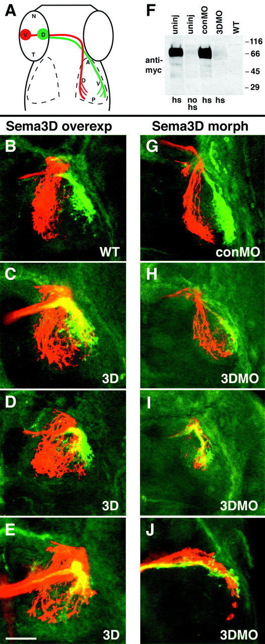
Retinotectal mapping by ventral RGC axons along the dorsoventral axis is perturbed by misexpression and MO knock-down of Sema3D. A, Schematic showing injection sites in retina and normal arborization locations. B-E, Confocal projections of 3 dpf embryos showing that ventral but not dorsal RGC axons extend aberrantly after misexpression of Sema3D. All are dorsal views (anterior is to the top) of whole-mounted embryos labeled with DiI (red) injection into ventral retina and DiA (green) injection into dorsal retina. Left eyes were injected, and right tectum is shown. Wild-type (B) and hsp70:sema3Dmyc transgenic (C-E) embryos, all heat induced at 36, 42, and 48 hpf and fixed at 3 d. Ventral RGC axons aberrantly extend into ventral tectum after Sema3D overexpression, but the dorsal RGC axons extend normally (C-E). Area of overlap between ventral and dorsal RGC axons is yellow. F, Western blot showing sema3D antisense MO knock-down of Sema3D protein expression in hsp70:sema3Dmyc embryos. Lanes 1-4 are hsp70:sema3Dmyc embryos. Lane 1, Uninjected, heat induced. Lane 2, Uninjected, not heat induced. Lane 3, conMO injected, heat induced. Lane 4, 3DMO injected, heat induced. Lane 5, Wild type. Blot is processed with anti-myc antibody. G-J, Confocal projections of 5 dpf embryos showing that ventral but not dorsal RGC axons map aberrantly in the tectum after Sema3D knock-down. Left eyes were injected, and right tectum is shown. G is conMO-injected embryo and H-J are 3DMO injected. Ventral RGC axons (red) extend aberrantly into ventral tectum after 3DMO injection, but dorsal RGC axons (green) extend into their normal target, the ventral tectum. Scale bar, 50 μm.
