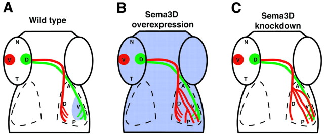
Figure 7.
Schematic summary of Sema3D manipulation results. Schematic dorsal views of wild-type (A), Sema3D-overexpressing (B), and Sema3D MO-injected (C) embryos. Sema3D expression is represented in blue. A, In wild-type embryos, Sema3D expression in the ventral tectum inhibits ventral retinal axons (red) from invading ventral tectum. B, When Sema3D is overexpressed uniformly, most ventral retinal axons extend correctly; however, their arbors also are spread into the ventral tectum. C, When Sema3D is knocked down, many ventral retinal axons extend into the ventral tectum, indicating the loss of an inhibitory cue there.

