
Figure 2.
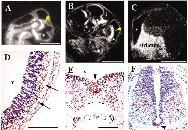
Embryonic FoxP2 mRNA (A–C) and protein (D–F) expression. Sagittal sections through stage 26 (A) and 34 (B) zebra finch embryos show expression in presumptive striatum (arrowheads) and presumptive dorsal thalamus (arrow). The heads face toward the right. C, Embryonic chicken brain (embryonic day 13) had strong expression in the developing striatum and also in the pallial and subpallial germinal ventricular zone, shown in a frontal right hemisection. The FoxP2 mRNA label appears white in dark-field illumination in A–C. D–F, FoxP2 expression in a stage 26 zebra finch embryo frontal sections. FoxP2 immunoreactivity is brown, and cresyl violet-stained cells are purple/blue. D, A prominent band of FoxP2-positive cells is visible among cresyl violet-stained neurons in the ventrolateral telencephalic vesicle. E, The floor plate at the rostral end of the mesencephalic vesicle (arrowhead) has many FoxP2-expressing cells that seem to disperse laterally (arrows). F, At limb levels of the spinal cord, floor plate neurons expressed FoxP2 (arrowhead), as did a population of neurons in ventral cord (arrows). Scale bars: A–C, 2 mm; D–F, 100 μm.