Figure 5.
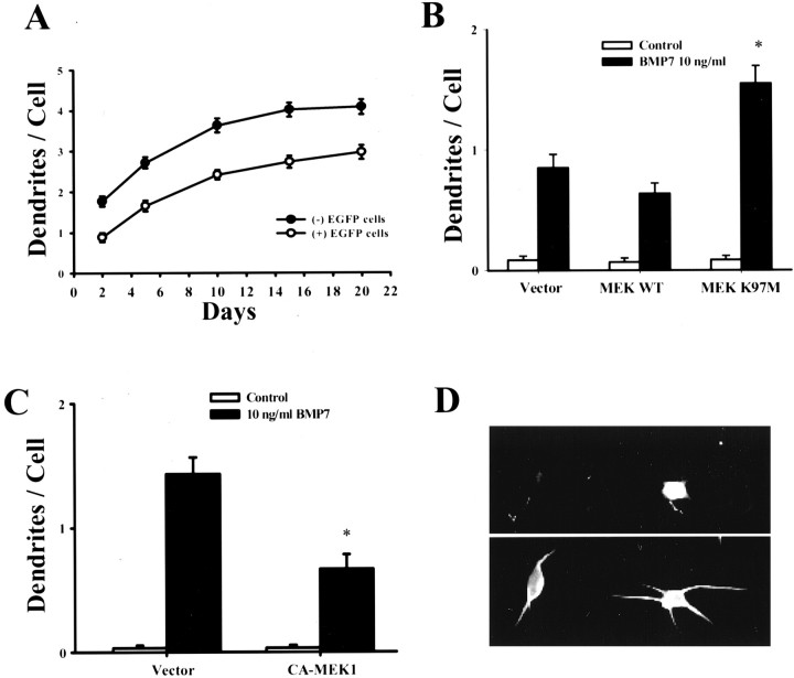
Dominant-negative MEK1 potentiates BMP-7-induced dendritic growth. For transfections, sympathetic neurons were plated at threefold higher density (∼30 cells/mm2) than in previous experiments. Under these conditions, BMP-7 (10 ng/ml) still induced dendritic growth, but the magnitude of the response was reduced (A). Neurons were cotransfected with plasmids containing EGFP and wild-type MEK1 (MEK1 WT) or dominant-negative mutant (MEK1 K97M) (B) or with a constitutively active mutant (C). Two days later, cells were treated with BMP-7 (10 ng/ml). On day 5, cellular morphology was assessed by immunostaining with an mAb to MAP2 (D, bottom; n ≥ 60 per group). Fluorescence micrograph of a nontransfected neuron and a neuron cotransfected with EGFP and dominant-negative MEK (MEK1 K97M) (D). Transfected cells were identified by expression of EGFP (D, top). *p < 0.05 versus BMP-7.

