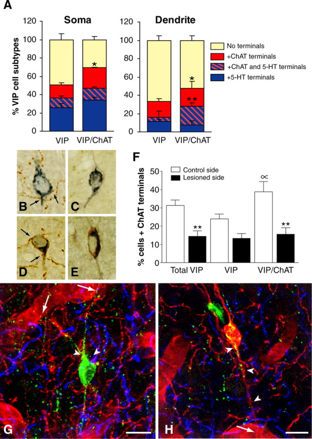
Figure 5.

Identification of subsets of VIP interneurons and their ACh and 5-HT afferents. A, In triple-labeled sections for VIP, ChAT, and 5-HT, comparable proportions of soma (∼40%) and dendrites (∼20%) from VIP and VIP/ChAT neurons received 5-HT afferents (blue box). In contrast, a larger proportion of VIP/ChAT than VIP neurons was contacted by ACh afferents (red box) on both cell soma (n = 216; p < 0.05) and dendrites (n = 186; p < 0.05), and a larger proportion of VIP/ChAT dendrites also received both types of afferents (**p < 0.01; hatched red and blue box) compared with VIP dendrites. B-F, Effect of unilateral basal forebrain lesion on the ACh afferents to VIP interneurons as evaluated in semithin sections labeled for VIP (SG kit; blue) and ChAT (DAB; brown). Quantitative analysis of VIP and VIP/ChAT neurons on the control (B, D; n = 476 cells) and lesioned (C, E; n = 496 cells) sides, respectively, showed a significant loss of ACh input after lesion of substantia innominata (F). Approximately 50% of the ACh innervation to the total VIP cell population (**p < 0.01) originated in the basal forebrain, with VIP/ChAT cells being significantly denervated (60%; **p < 0.01) but not the VIP (44%; NS) cells. Also, the analysis on the control side confirmed that VIP/ChAT cells received more ACh afferents than VIP cells (∝; p < 0.05). G, Quadruple immunofluorescence of a perivascular VIP neuron (green; Cy2) contacting a neighboring blood vessel (arrows; laminin-immunodetected with Cy3, red) and receiving both ACh (ChAT and Cy3; red) and 5-HT (5-HT and Cy5; blue) afferent terminals on its cell body or proximal dendrites (arrowheads). H, Quadruple immunofluorescence as in G but showing a VIP/ChAT interneuron (red/yellow) in contact with a blood vessel (arrow) and receiving dual ACh (red) and 5-HT (blue) innervations (arrowheads). Scale bars, 10 μm.
