
Figure 5.
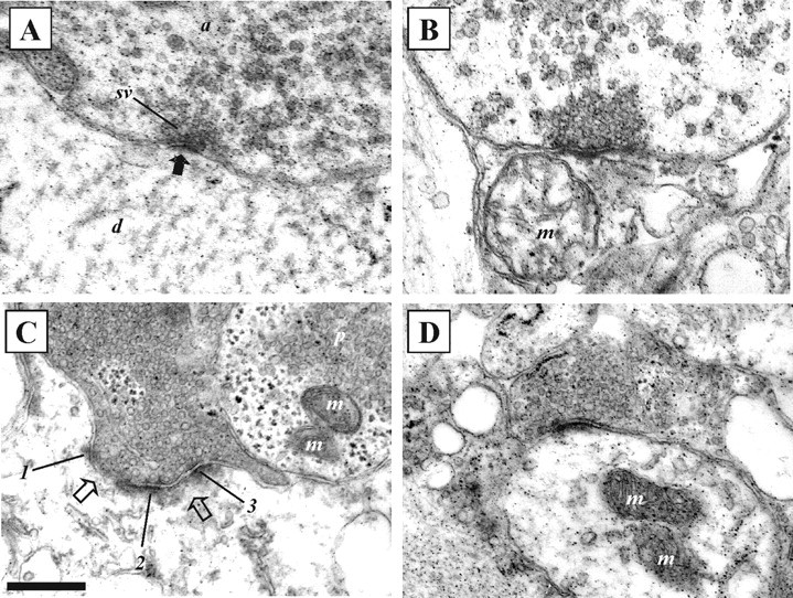
Electron micrographs showing examples of synaptic morphology before and at different times after substance P application. A, Example of an asymmetric synapse with round vesicles in a spinal cord that had not been exposed to substance P. The solid black arrow indicates the PSD. B, An asymmetric synapse with round vesicles 30 min after substance P application. C, An asymmetric synapse with two perforations (open arrows) and round vesicles 3 hr after substance P application. Numbers 1-3 indicate regions of the PSD. A neighboring axon contains pleomorphic vesicles (p; vesicles with irregular shapes and sizes). D, An asymmetric synapse with round vesicles 5 hr after substance P application. a, Axon; d, dendrite; m, mitochondria; sv, synaptic vesicles. Scale bar, 250 nm.