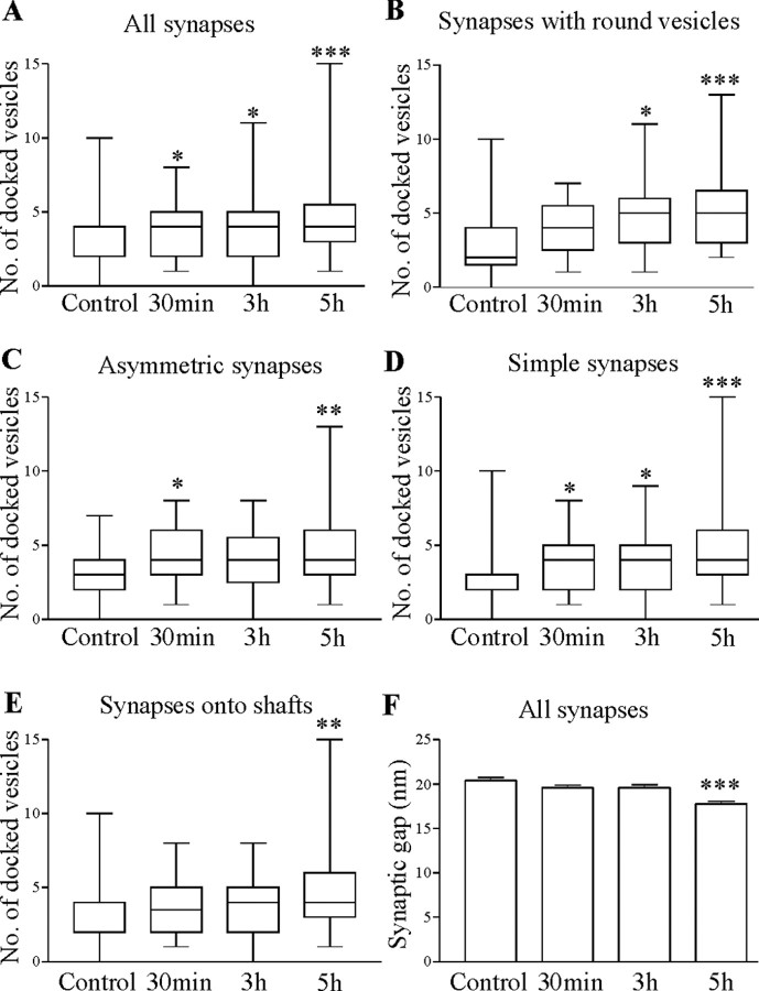
Figure 6.
Substance P increased the number of docked vesicles and reduced the synaptic gap. A, Graph showing the number of docked vesicles at all synapses at different times after substance P application. Graphs showing the number of docked vesicles at synapses with round vesicles (B), asymmetric synapses (C), simple synapses (D), and synapses made onto dendritic shafts (E). F, Graph showing the distance across the synaptic gap at different times after substance P application. A-E, Pooled data: control, n = 4 animals; 30 min, n = 4 animals; 3 hr, n = 3 animals; 5 hr, n = 4 animals. F, Pooled data: control, n = 5 animals; 30 min, n = 4 animals; 3 hr, n = 4 animals; 5 hr, n = 4 animals. *p < 0.05; **p < 0.01; ***p < 0.001.

