
Figure 1.
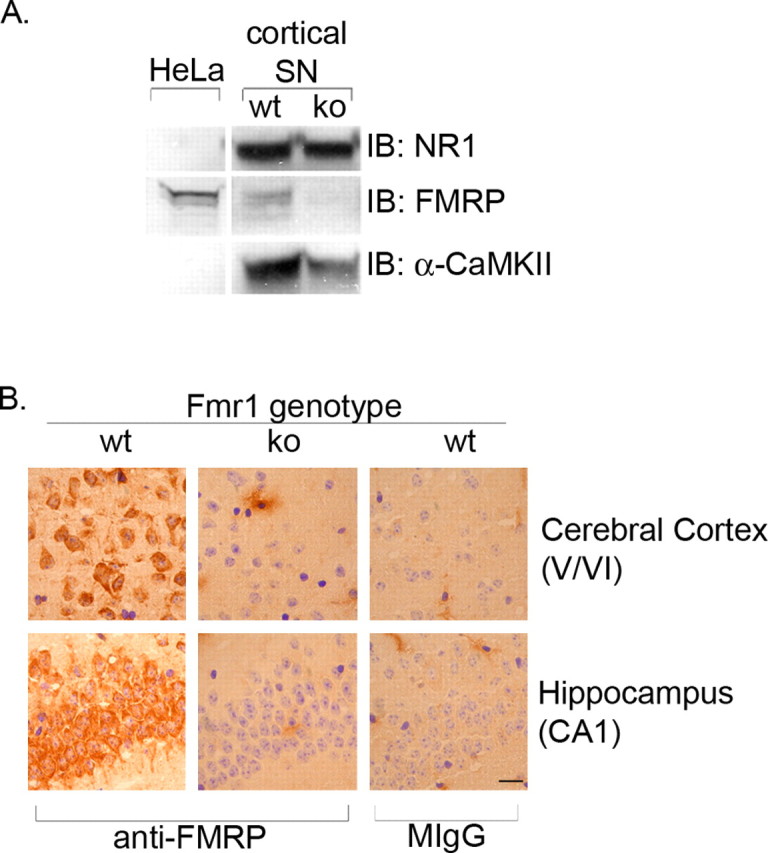
Antibody 2F5-1 is FMRP specific. A, Synaptoneurosome (SN) fractions were prepared from the cortex of wild-type (wt) and Fmr1 null mice (ko) and probed for FMRP, NR1, and α-CaMKII expression by Western blot. HeLa cell extracts are used as a positive control for FMRP expression. B, Sagittal sections from wild-type and Fmr1 knock-out mice were immunostained with FMRP or normal mouse IgG (MIgG; control). FMRP-positive cells are visible in the cerebral cortex and hippocampus of wild-type mice, but no FMRP-positive cells are detected in Fmr1 null mice. Scale bar, 10 μm.