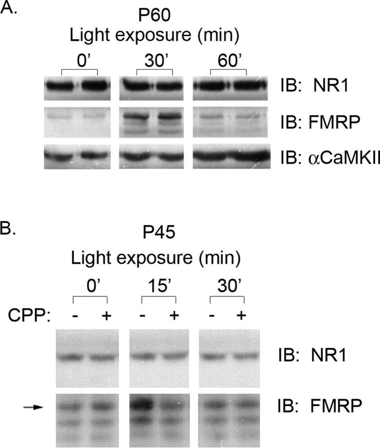
Figure 3.
Visual experience induces a transient increase in FMRP levels in synaptoneurosome fractions from visual cortex. A, P60 DR rats were LE for 0, 30, or 60 min. Synaptoneurosome fractions were prepared from the visual cortices and probed for FMRP, NR1, and α-CaMKII expression by Western blot. In DR animals of this age (P60), FMRP levels peak at 30 min and return to baseline by 60 min. In contrast, α-CaMKII levels are elevated at 30 min and further increased at 60 min. Each lane shows synaptoneurosome-enriched fractions prepared from the visual cortex of an individual rat. B, The upregulation of FMRP induced by visual experience is NMDA receptor dependent. P45 dark-reared rats were exposed to light for the indicated times. The increased expression of FMRP observed at 15 min of light exposure is blocked by systemic administration of the NMDA receptor antagonist CPP (10 mg/kg). Systemic administration of MPEP (10-20 mg/kg) had no effect on FMRP expression (data not shown).

