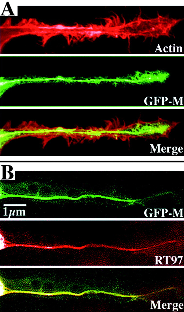
Figure 1.

Axonal cytoskeletons display an array of filamentous actin and NFs and incorporate GFP-tagged NF subunits into the endogenous NF network. The panels present axonal neurites of two cells transiently transfected 24 hr previously and then fixed and extracted with Triton X-100. The top cell (A) was reacted with rhodamine-conjugated phalloidin to visualize filamentous actin, and the bottom cell (B) was reacted with a monoclonal antibody (RT97) directed against a phosphorylated NF epitope. Note the peripheral concentration of filamentous actin. Note that both actin and GFP fluorescence are present along the entire length of the axonal neurite; however, actin is concentrated along the periphery, whereas GFP-M is concentrated along the central aspect of the neurite. This central concentration corresponds to the bundle of phospho-NFs (Yabe et al., 2001a, b) as confirmed in the bottom neurite by preferential labeling of this bundle by RT97. Also note the colocalization of GFP-tagged NF subunits with phospho-NFs in the merged image, indicating their incorporation into the NF cytoskeletal network.
