Figure 11.

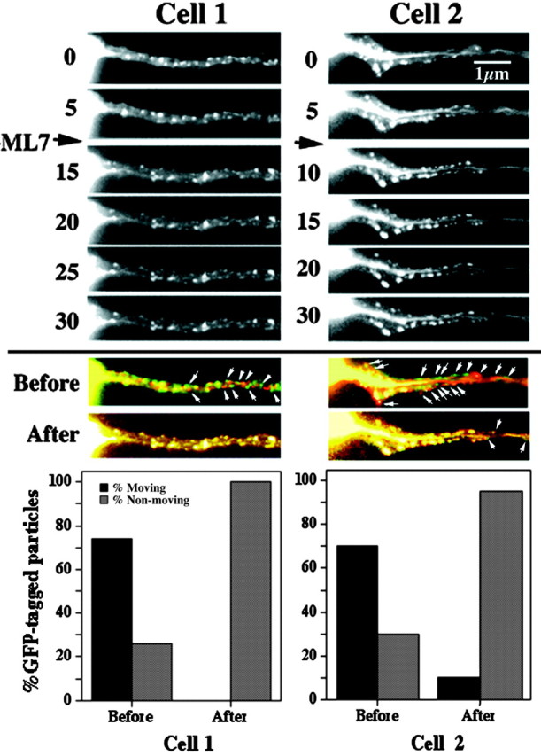
ML-7 inhibits bidirectional transport of GFP-tagged NFs and punctate structures. Sequential images captured at the indicated intervals of axonal neurites of two transfected cells before and after the addition (arrow) of 300 nm ML-7 are shown. Small arrows denote representative GFP-tagged structures. As described in Materials and Methods, two sequential images before the addition of ML-7 were false-colored with either red and green, and a merged image was created (Before); the same procedure was performed for two sequential images after the addition of cytochalasin B (After). Particles that had not translocated during the interval between the capture of images displayed a merged orange-yellow fluorescence, whereas those that had undergone translocation displayed either red or green fluorescence. The accompanying graphs present the percentage of particles displaying either red or green fluorescence versus those displaying orange-yellow fluorescence. Note that ML-7 reduced the percentage of translocating particles. A video sequence of the effect of ML-7 on GFP-tagged particle translocation is presented as supplemental material (available at www.jneurosci.org). The depicted cells correspond to those presented in Figure 2 before the addition of ML-7.
