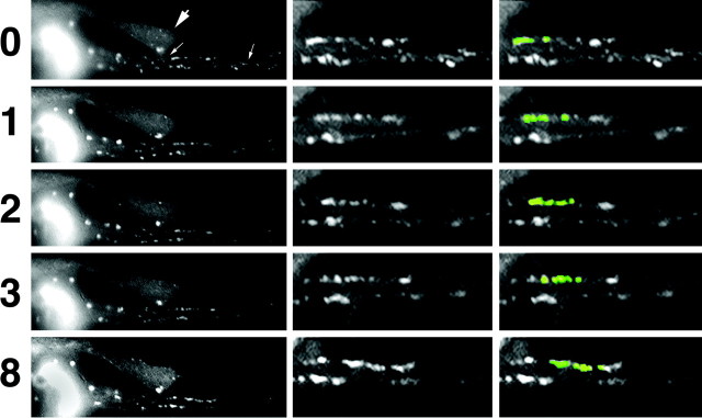
Figure 2.
GFP-tagged NFs and punctate structures undergo bidirectional translocation along the inner plasma membrane of axonal neurites. Images captured sequentially under fluorescein optics at the indicated intervals (in minutes) are presented. The left panels present the axonal neurites of cells transfected with GFP-M. Small arrows denote the region of a cell in which GFP-tagged subunits have not yet incorporated throughout the entire endogenous NF network, which facilitates observation of particles undergoing translocation along the membrane; the region denoted by arrows is presented at higher magnification in the middle panels and after false coloration of select particles in the right panels. The large arrow to the left denotes the neurite of a second transfected cell in which GFP-tagged subunits previously have incorporated throughout the axonal NF network; note that this obscures the localization and movement of individual GFP-tagged structures during the analysis of sequential images. False color to highlight the video sequence corresponding to these images is presented in QuickTime format (supplemental material, available at www.jneurosci.org).

