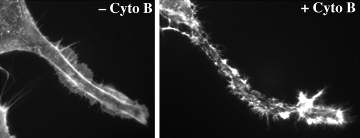
Figure 3.
Cytochalsin B treatment disrupts the submembrane actin network. The panels present portions of perikarya and axonal neurites of cells before (left) and after (right) 2 hr of treatment with 5 μm cytochalasin B. Note the continuous distribution of actin-rich filamentous profiles in untreated cells and their perturbation after cytochalasin B treatment.

