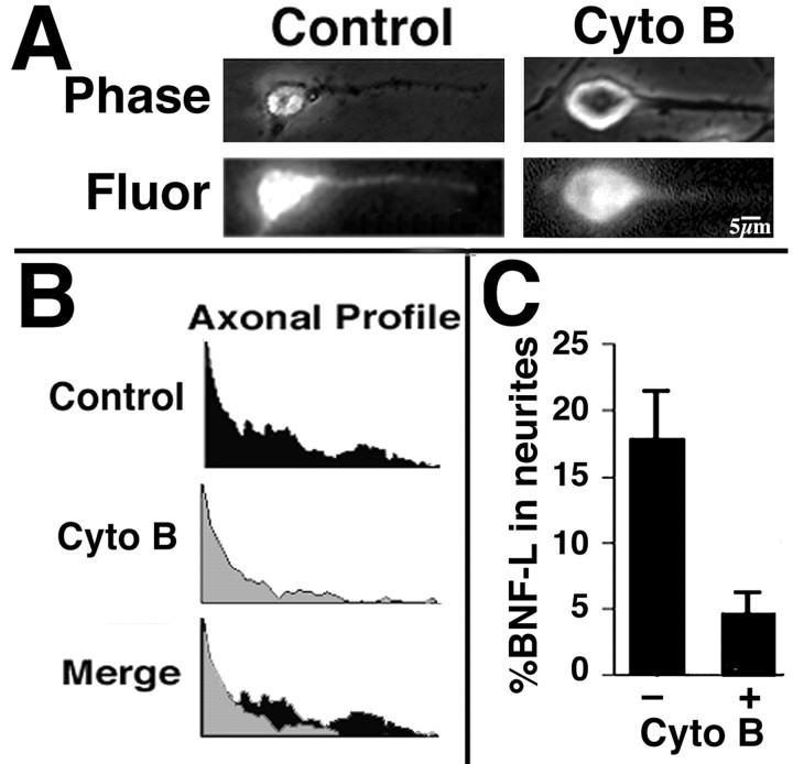
Figure 4.
Cytochalsin B treatment inhibits transport of biotinylated NF subunits into axonal neurites. A, The panels present phase-contrast and corresponding epifluorescent images of representative cells microinjected with bNF-L or tracer alone and then fixed and immunostained for biotin at 1 hr after injection; alternate cultures received 5 μm cytochalasin B immediately after injection. B, We visualized the distribution of bNF-L (Axonal Profile) within cytochalasin B-treated and cytochalasin B-untreated axonal neurites for the presented images by encircling the axonal neurite from hillock to growth cone and invoking the plot profile function of NIH Image. Overlaying of plot profiles (Merge) from untreated and treated neurites highlights that both vinblastine and cytochalasin B inhibited translocation of bNF-L into neurites. C, The accompanying bar graph presents the percentage of bNF-L that had translocated into neurites within 1 hr after microinjection (mean ± SD; n ≥ 3 cells for each condition from ≥3 separate experiments for a total of ≥9 individual injected cells), calculated by dividing the fluorescent intensity within neurites by that within the neurite plus the corresponding cell body. Note that cytochalasin B markedly reduces the transport of biotinylated NF subunits into neurites.

