Figure 5.

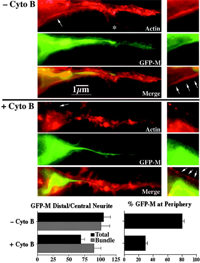
Cytochalasin B inhibits the distribution of GFP-tagged NF subunits. The panels present epifluorescent images of representative cells transfected with GFP-M and reacted with rhodamine-phalloidin before (-Cyto B) and after (+Cyto B) treatment with cytochalasin B. Note that cytochalasin B reduced the total level of GFP-M within the distalmost region of the neurite but did not seem to reduce GFP-M levels within the central bundle. Also note the absence of GFP-tagged NFs from an actin-rich filopodium (asterisk), confirming discrete staining. The accompanying left graph presents densitometric analyses of the ratio of total and bundle-associated GFP-M in the distal third of axonal neurites versus the central third; these densitometric analyses confirm the visual impression that cytochalasin B reduced overall GFP-M, but not bundled GFP-M, with distal axonal regions. The panels on the right present higher-magnification images of the region denoted by a small arrow in the image on the left. Arrows within the higher-magnification images indicate the peripheral region of the cell. The accompanying right graph presents densitometric analyses of the ratio of GFP-M at the periphery versus within central regions before and after cytochalasin B treatment. Note that cytochalasin B reduced the localization of GFP-M along the periphery.
