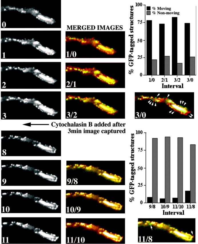
Figure 6.

Cytochalasin B reduces translocation of GFP-tagged NFs and punctate structures. The panels present images captured sequentially at the indicated intervals (in minutes) of the distal region of a neurite of a cell transfected 24 hr previously with GFP-M before and after the addition of cytochalasin B. To facilitate comparison of particle translocation, we false-colored sequential images with either red or green and created a merged image for each interval; units in merged images correspond to the time of capture of individual images. Particles that had not translocated during the interval between the capture of images displayed a merged orange-yellow fluorescence, whereas those that had undergone translocation displayed either red or green fluorescence. Arrows in the final merges before and after the addition of cytochalasin B denote representative GFP-tagged particles. The accompanying graphs present the percentage of particles displaying either red or green fluorescence versus those displaying orange-yellow fluorescence for each interval before and after the addition of cytochalasin. Note that cytochalasin B markedly reduced the percentage of translocating particles.
