
Figure 1.
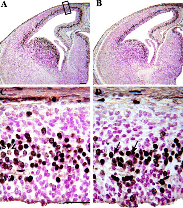
Photomicrographs illustrating BrdU labeling in E14 control (A, C) and nestin/IGF-I Tg (B, D) embryos 0.5 hr after injection of a timed-pregnant dam. Counts of BrdU-labeled and unlabeled cells were conducted over the dorsomedial cerebral wall (A, black rectangle). BrdU-labeled cells were identified by the accumulation of brown reaction product in cell bodies (D, arrows). Unlabeled cells appear pink because of the use of a basic fuchsin counterstain. As illustrated in the low-power photomicrographs, Tg embryos (B) were not visually distinguishable from controls (A) on E14. C and D illustrate the structure of the cerebral wall from the ventricular surface to the pia at 9:30 A.M. on E14. The cerebral wall consisted mainly of the VZ and SVZ in both control (C) and Tg (D) embryos at this time, and a morphologically distinguishable cortical plate was not evident in either group. The height of the VZ and the total cerebral wall were similar in Tg and control embryos. Scale bars: A, 100 μm; C, 30 μm.