
Figure 5.
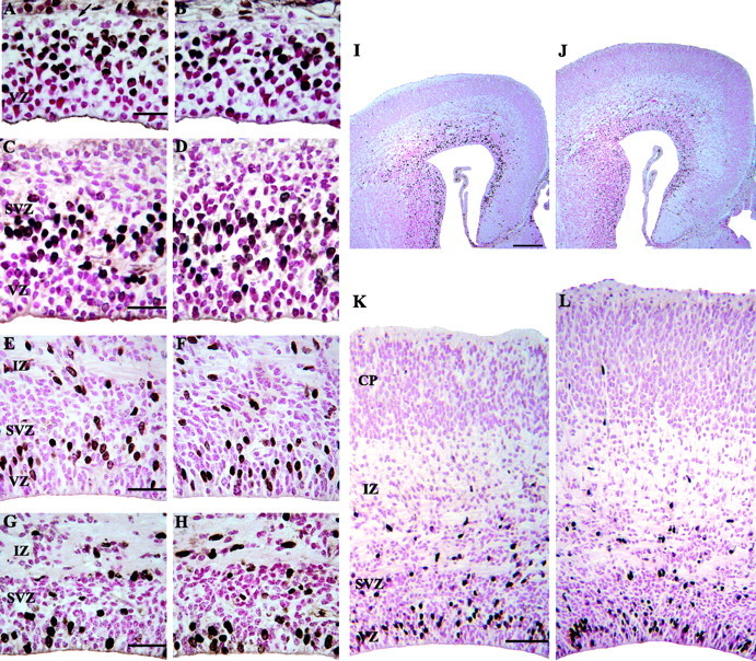
Representative photomicrographs of the dorsomedial cerebral wall at E11, E14, E17, and E18 in control (A, C, E, G) and nestin/IGF-I Tg (B, D, F, H) embryos. Embryos were labeled with BrdU and killed at 1 hr after injection. At E11, the cerebral wall consisted of the VZ and an adjacent zone of postmitotic cells (A, arrow). By E14, the SVZ was apparent adjacent to the VZ (C, D). The height of the VZ did not differ between Tg (D) and control (C) embryos. A distinct zone of BrdU-labeled cells was apparent in the VZ, indicating that progenitors were actively proliferating. At E17, the VZ was reduced in height compared with E14 embryos in both control (E) and Tg (F) embryos, but a zone of BrdU-labeled cells in S phase was still evident. Therefore, neurogenesis was still occurring at E17, but the reduced height of the VZ suggested that this process was nearing completion. Increased neuron output from the VZ was apparent in control (I, K) and Tg (J, L) embryos at E17, because the IZ and CP were increased in size by this age. By E18, a distinct VZ was not apparent in either control (G) or Tg (H) embryos. BrdU labeling showed random staining in the cells adjacent to the ventricular surface, which were likely ependymal cells. Nestin/IGF-I Tg embryos were visibly larger than control embryos by E17 (I, control; J, Tg). Closer examination of histological sections showed that the cortical plate was much larger in Tg embryos (L) compared with controls (K). Scale bars: A, C, E, G, 20 μm; I, 100 μm; K, 70 μm.