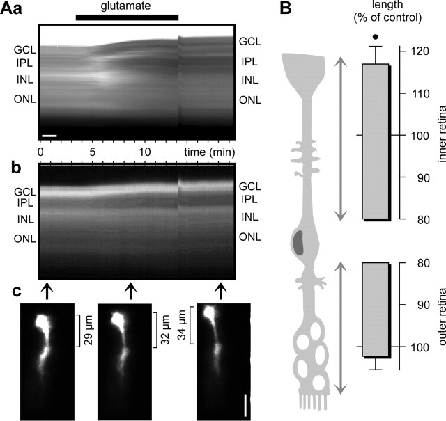
Figure 2.
Glutamate (1 mm) evokes thickening of retinal slices and elongation of single dye-filled glial (Müller) cells. A, Example of one slice. Aa, Intrinsic optical signals in dependence on the recording time. Agonist application is indicated by the thick bar. There is an artifact at the time of removal of the agonist. Ab, Time dependence of the fluorescence signal of one Lucifer yellow-filled glial (Müller) cell. Ac, Examples of fluorescence records of the dye-filled cell at three different time points indicated by the arrows in Ab. The distance between soma and end foot of the cell is indicated. Scale bars, 20 μm. B, Lengths of the outer and inner retinal parts of fluorescence dye-filled glial (Müller) cells, measured after 10 min of glutamate exposure. The data were obtained in four independent experiments and are expressed as percentage of control (100%). •p < 0.05.

