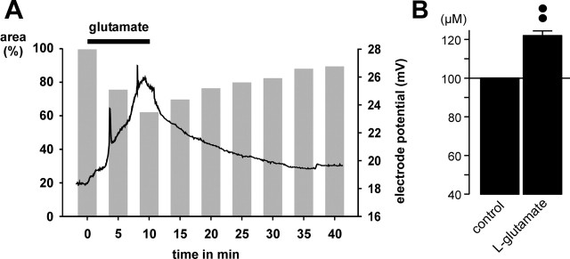
Figure 6.
Glutamate application causes a decline of the volume of the extracellular space in the IPL of the guinea pig retina. The volume was measured by using TMA+-containing bath solution and TMA+-sensitive microelectrodes. A, The trace shows that bath application of glutamate (1 mm) results in a reversible increase of the electrode potential, which reflects an increase of the extracellular TMA+ concentration in the IPL. The gray bars show the mean time-dependent changes of the cross-sectional area of glial cell profiles. The black bar indicates agonist exposure. B, Mean (±SEM) TMA+ concentration in the IPL before (control) and 10 min after the onset of glutamate (1 mm) exposure (n = 5). ••p < 0.01.

