Figure 7.
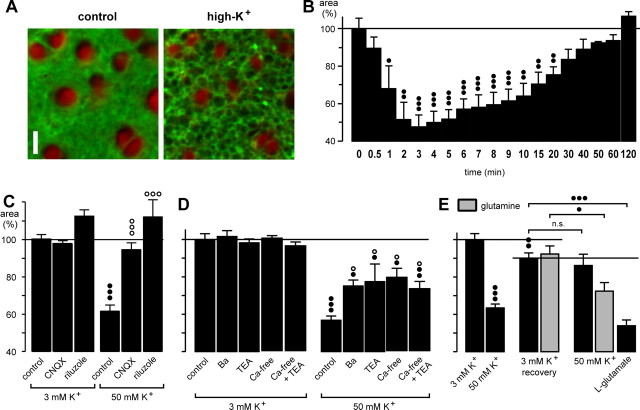
High extracellular K+ evokes transient morphological alterations in the guinea pig retina that are caused by stimulation of endogenous glutamate release. A, Elevation of the K+ concentration in the bath solution from 3 to 50 mm caused a decrease of the cross-sectional areas of glial cell profiles in the IPL (recorded before and after 5 min of high-K+ exposure). Cross sections through Müller cell processes were red-stained by Mitotracker Deep Red, and cell membranes were stained green by FM 1-43. Scale bar, 5 μm. B, Time dependence of the high-K+ (50 mm) effect. C, The effect of high K+ was inhibited by coapplication of the AMPA-kainate receptor blocker CNQX (50 μm) and of the glutamate release blocker riluzole (500 μm), respectively. D, The effect of high K+ was partially inhibited by coapplication of Ba2+ (1 mm) or TEA (10 mm) and in the presence of a Ca2+-free extracellular solution (with or without 10 mm TEA). E, The presence of glutamine in the extracellular solution partially restored the effect of high K+ on the glial morphology during a second application. The whole mounts were superfused for 10 min with a high-K+ (50 mm) solution that decreased the thickness of glial cell profiles by ∼40% (left). Subsequently, the high-K+ solution was washed out for 45 min in the absence or presence of glutamine (250 μm) (recovery; middle), and then the high-K+ solution was again applied for 10 min (right). As control, glutamate (1 mm) was tested. The cross-sectional areas of the glial cell profiles in the IPL are given as percentage of control (100%). In C and D, the effects were measured after 10 min exposures. Data were obtained in 3-17 independent experiments. Significant effect versus control: •p < 0.05; ••p < 0.01; •••p < 0.001. Significant effects of the blocker: ○p < 0.05; ○○○p < 0.001.

